
Cruciate Surgery & Treatment
Cruciate disease is a debilitating condition that affects dogs, but early recognition can make a significant difference in your pet’s quality of life. As a pet owner, educating yourself on the signs can help you seek timely treatment. We understand how heartbreaking it is to see a beloved pet in pain, which is why our experienced veterinarians are here to help. With expert diagnosis and treatment, we are dedicated to restoring your dog’s health and happiness.
What is Cruciate Disease?
Cruciate disease involves trauma, injury or degeneration of the cranial cruciate ligament. The cranial cruciate ligament is an important structure that is responsible for stability and prevention of excessive rotation of the knee or stifle joint. When this ligament is injured or ruptured, it results in joint instability, leading to lameness, pain and joint swelling. The disease is often bilateral, meaning it affects both stifles with an incident rate between 22-54%.
Which Breeds Are Prone to Cruciate Disease?
Cruciate disease is the most common cause of lameness in the hindlimbs of dogs. Middle-large breed dogs are mainly affected such as Labradors, Rottweilers, Newfoundlands and Staffordshire Terriers.
Acute traumatic injury to the cranial cruciate ligament is rare and often occurs in young dogs. This injury often occurs due to abrupt hyperextension or over-rotation of the knee joint, such as when chasing the ball in the park.
More commonly, dogs suffer from a chronic or degenerative form of cruciate disease, which results from the slow weakening of the ligament over time. As the ligament fibres begin to fray or weaken, this will eventually lead to partial or complete rupture of the ligament. Dogs between 5-7 years old are mostly affected. These dogs often have a chronic history of intermittent lameness but can have acute flare-ups or suffer from a traumatic event that can speed up the process of complete ligament rupture.
What Causes Cruciate Disease in Dogs?
The cause of chronic cruciate disease is not completely understood but can be related to anatomical confirmation, breed predisposition, obesity, hormonal and auto-immune influences. There is a higher incidence of females compared to males that are affected.
In 50% of cases of cruciate disease, damage will also be identified within the medial meniscus, which is a cartilage pad that sits on the inner aspect of the knee joint.
What Are the Clinical Signs of Cruciate Disease?
- Limping or lameness
- Pain when attempting to stay in a sitting position. This can result in dogs keeping their leg in an extended or straightened position when sitting.
- Pain when flexing or extension the knee/stifle joint.
- Swelling of the knee/stifle joint.
- Crepitus or clicking when manipulating the knee joint.
- Muscle loss or atrophy.
- Thickening of the joint capsule on the medial aspect of the knee causing the formation of a “medial buttress”.
- Positive cranial draw or tibial compression test.
How is Cruciate Disease Diagnosed?
- Clinical history.
- Conscious musculoskeletal and orthopaedic exam.
- Examination of the stifle joint under sedation.
- Radiographs or x-rays of the affected stifle.
If cruciate disease is suspected, our veterinarians will recommend booking your pet for examination and radiographs of the affected joint under sedation. This is to allow us to perform a test called the cranial draw sign, which is used to identify a ruptured cruciate ligament. This test is often not successfully performed in a conscious patient as they are often too painful and tense for the test to be accurate.
The cranial draw sign test involves placing one hand above and below the stifle joint and attempting to move the tibia or shin bone forward. If the ligament is ruptured, the tibia bone will move excessively forward and feel unstable, which is a positive cranial draw sign. An intact ligament will prevent the tibia from moving forward.

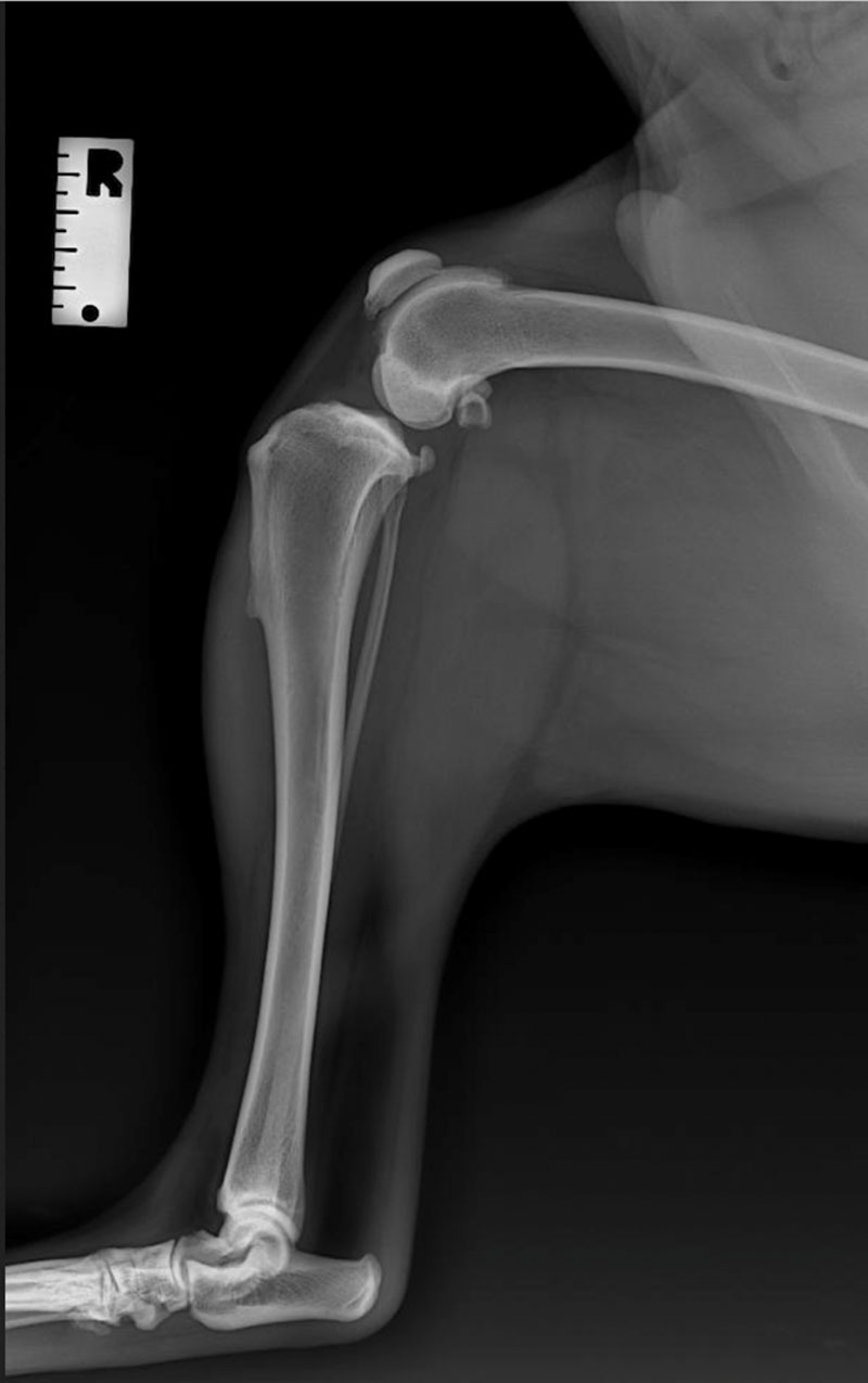
Radiographs can also highlight evidence that your dog has cruciate disease. Common findings on X-rays are swelling or effusion within the stifle joint and early osteoarthritis changes.
What is Involved in Cruciate Surgery?

There is one main consideration when it comes to cruciate surgery, and that is the weight.
For Dogs That Weigh Less Than 15kg
The De-Angelis procedure is performed in dogs <15 kg. It is known as an extracapsular repair technique which involves placement of a synthetic suture on the outside of the knee or stifle joint to mimic the pathway of the cruciate ligament.
During the procedure, an arthrotomy is performed, which involves opening the knee or stifle joint to assess the cruciate ligament. The damaged cruciate ligament is removed via sharp dissection, and the medial meniscus is assessed for damage. If the medial meniscus is found to have a tear, it will also be transected and removed via a technique called a meniscectomy. The joint is then copiously lavaged or flushed with sterile saline before closure.
A non-absorbable suture is then anchored behind a structure called the lateral fabella on the outside of the knee joint. The free suture ends are then buried underneath a muscle called the cranial tibial muscle and passed beneath the patella ligament through bone tunnels created via a drill in the proximal tibial bone. The suture ends are then tied on the opposite or medial side of the stifle joint. The sutures are tightened, and joint stability is confirmed by a negative cranial draw test.
The overlying soft tissue structures are then closed followed by the skin. An imbrication technique, which involves removing a portion of soft tissue during closure, is often performed to help keep the suture more secure.
This technique aims to stabilise the joint long enough until scar tissue or fibrosis of the knee creates permanent stability.
For Dogs That Weigh More Than 15kg
The TPLO (Tibial Plateau Levelling Osteotomy) procedure is an advanced surgical technique performed in dogs >15 kg. Due to the complexity and equipment involved in the procedure is performed via a specialist orthopaedic surgeon who visits Berwick Clyde Vet for the day. Currently, we use Dr Ralph Webster from Better Veterinary Referral Services.
An arthroscope is a non-invasive technique which involves placement of a camera device inside the knee joint to evaluate the cruciate ligament and the meniscus. A shaving device is then inserted into the joint to debride the remnants of the damaged ligament. If the meniscus is damaged, a meniscectomy will be performed. The joint is then flushed with copious amounts of sterile saline and the incisions created to place instruments into the joint are sutured closed. An arthroscopy will be conducted prior to the TPLO procedure.
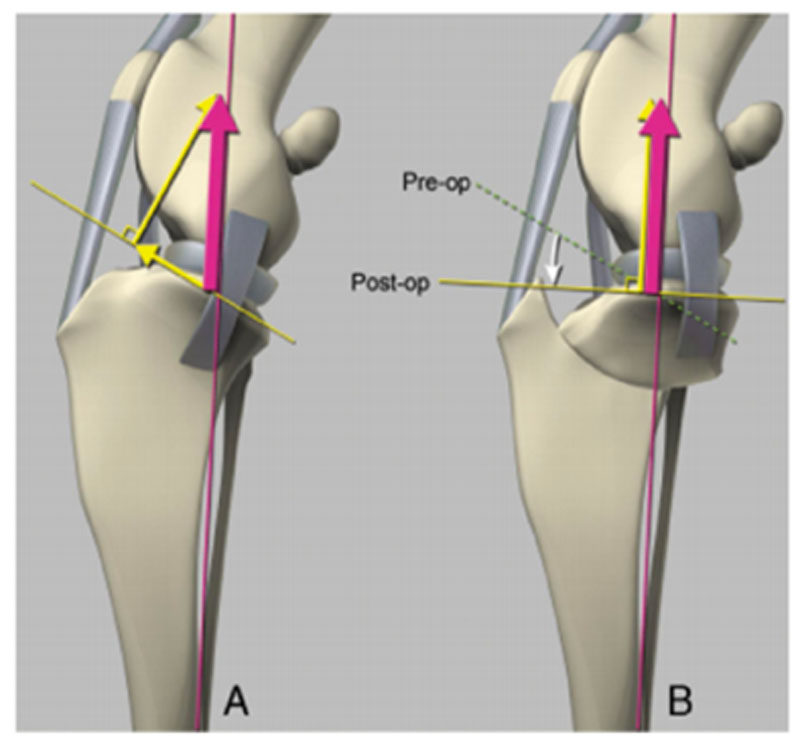
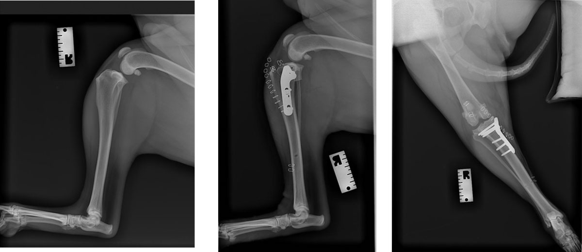
The TPLO technique involves changing the angulation of the stifle joint and flattening the tibial plateau. The tibial plateau is approximately 24 degrees in the average dog. When a dog places pressure on its hindlimb or a weight-bearing force is directed down the leg and down the tibial plateau. When the cruciate ligament is ruptured, the tibial bone pushes forward. During a TPLO the tibial plateau angle is reduced to 6 degrees by performing an osteotomy, which involves cutting and rotating the tibial bone. The bone is stabilised or secured in place with a plate and bone screws. These screws are not routinely removed. By reducing the tibial plateau angle, the tibial bone is prevented from moving forward during weight bearing, improving stability of the knee.
Are there complications associated with the De-Angelis or TPLO procedure?
- Post-operative infection – reported in 5% of cases.
- Implant failure, suture breakage or slippage – reported in 1-5% of cases.
- It is common for the De-Angelis suture to fail or become loose overtime but often dogs will still have good clinical function of the limb with minimal lameness. However, if the suture breaks completely, this will result in instability and pain, which is why revision surgery is often required.
- Meniscal injury – reported in 3-8% of cases post TPLO.
- Wound breakdown.
What is Involved in the Recovery for Cruciate Surgery?
Rest
Following the De Angelis or TPLO procedure it is recommended that your dog is kept crate or cage rested for 8 weeks post-surgery. If your pet is not comfortable resting in a crate, you can set them up in a small room or area in the house, such as the bathroom or laundry. You can apply an icepack to the surgical site during the first 48-72 hours post-surgery to help alleviate swelling and inflammation. Some dogs will not tolerate ice packing; therefore short and more frequent sessions are ideal e.g. icepack for 3-5 minutes 3-4 times daily.
The First Two Weeks After Operation
The most critical period is the first 2 weeks following surgery, as this is when there is the highest risk of implant failure. Strict rest and only short-leashed walks to the toilet are permitted in the first 10-14 days of the recovery period.
When to Re-introduce Exercise
After this, you can begin to slowly and incrementally increase the level of exercise your dog can tolerate. It is advised to start with 5–10-minute leashed walks once to twice daily and increase the length of walks by 5-10 minutes each week. By 8 weeks post-operatively, your dog should be able to tolerate close to their normal exercise levels pre-surgery.
Physiotherapy
Physiotherapy is recommended to help strengthen the surgical limb and to fasten recovery. At home exercises such as gentle massage and passive range of motion can be started as early as 3 days post-surgery. Our vets may also recommend that your dog seeks guidance and sessions with a qualified canine physiotherapist at facilities such as Dogs in Motion and Aquapaws. These facilities also provide hydrotherapy options which involve walking exercises on an underwater treadmill to again promote muscular strengthening.
Medication
Your dog will be sent home with adequate pain-relief, including non-steroid anti-inflammatory medication and opioid medication to keep them pain-free and comfortable during recovery. We advise that your dog wears an Elizabethan collar until the surgical incision is healed, which can take up to 10-14 days.
Long-term Management Plan
We will also commonly organise a long-term osteoarthritis management plan with your pet following the procedure. All dogs that have cruciate disease will, unfortunately, develop osteoarthritis in the future. We will discuss preventative measures such as starting osteoarthritis injections (e.g. zydax, beransa) and supplements (e.g. 4 cyte).
If your dog has undergone a TPLO procedure, they will be required to return to the clinic 8 weeks post-operatively for sedation and x-rays to check healing of the osteotomy site before they can return to normal off-leash activity.
Why Choose Us?
At Berwick Clyde Vet, we have a wealth of experience performing cruciate surgeries. We look after your pets as if they were our own. Our support for you and your pets does not end as you walk out the door; we are here for you every step of the way until your pet fully recovers.
If you would like to learn more about cruciate surgery for your pet, our friendly team is more than happy to help.